Affiliation: School of Medicine, Institute for Biogenesis Research
Position: Associate Professor
Degree: PhD (University of Toronto, Canada)
Phone: (808) 692 1417
Fax: (808) 692 1962
Email: vernadet@hawaii.edu
Address: 651 Ilalo St., Biosciences Building, Room 163J/H, Honolulu, HI 96813
Go to Vernadeth Alarcon's webpage
Preimplantation embryo, Cell lineage, Cell polarity, Developmental toxicants
Mammals, such as ourselves, are unique among other organisms in that our embryonic phase needs to implant into the mother's uterus and create the placenta to allow further development. To achieve such unique style of development, we have evolved to generate a special type of cells, called the trophectoderm, during the very early stages of embryo development (Figures 1 and 2). Trophectoderm is dedicated for implantation and placenta formation. In fact, the first and critical decision that embryonic cells must make after fertilization is whether they commit to become part of the future body or the trophectoderm, i.e., the precursor of placenta. This first decision needs to be controlled very carefully in a balanced manner because embryos cannot survive or develop further if one cell type (either "future body cells", called the inner cell mass, or "future placenta cells", the trophectoderm) is not generated sufficiently.
Our lab's research goal is to uncover the molecular mechanisms that control this early cell type decision-making, using mouse embryos as well as mouse and human cell lines. We have identified candidate genes, including cell polarity regulators (e.g., Alarcon 2010; Kono et al. 2014), and we are testing their roles in the embryo. We employ various experimental approaches, such as in vitro embryo culture, microinjection of the embryo, chimera production, embryo transfer into surrogate mother's reproductive tract, and cell and molecular biology techniques. Understanding the mechanisms of cell type decision-making in the early embryo has applications in the treatment of infertility, especially in ART (assisted reproductive technologies). Our findings have potential to serve as a basis for improving in vitro culture conditions of human embryos and for identifying healthy embryos for uterine transfer to produce successful pregnancies.
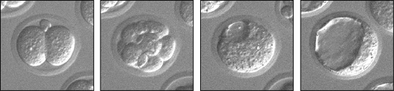
Figure 1: A preimplantation mouse embryo developing over time (left to right): 2-cell, 8-cell, early blastocyst, and late blastocyst. In the late blastocyst, cells have committed to the trophectoderm (future placenta) and inner cell mass (future body) lineages. Live development in vitro was recorded for three days, using time-lapse video microscopy.

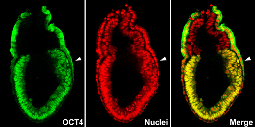
Figure 2: Immunofluorescently stained blastocysts, showing expression of transcription factors essential for cell-type commitment. (A) OCT4 (green) protein is localized in inner cell mass. (B) CDX2 (yellow) protein is localized in trophectoderm. Nuclei are stained red with propidium iodide. Images were taken using confocal microscopy.
 Richard Allsopp
Affiliation: School of Medicine, Institute for Biogenesis Research
Position: Professor
Degree: PhD
Phone: (808) 692 1412
Fax: (808) 692 1951
Email: allsopp@hawaii.edu
Address: 651 Ilalo St., BSB 163B, Honolulu, HI 96813
Analysis of the role of telomerase in stem cells and human diseases.
Telomerase is required to maintain telomeres, the protective cap at the end of chromosomes, in all eukaryotic cells. In mammals, including humans, some somatic cells in adults lack telomerase, and as a consequence, telomeres gradually short during cell division and as a function of age. Stem cells are required to replenish dead or damaged cells throughout life, and therefore telomerase is thought to play an important role in stem cell survival. While telomerase is indeed detectable in many types of human stem cells (unlike mature somatic cells), in some cases, such as in the blood, there isn't sufficient telomerase to maintain telomere length as these cells divide, and as a consequence, telomeres shorten in all blood cells during aging. On the other hand, male germ cells do have sufficient telomerase to thwart telomere loss during aging. one of the primary goals in my lab is to get a better understanding as to how telomerase activity levels are regulated in different types of stem cells. Recently, we have performed a screen for transcriptional regulators in embryonic stem cells and found that the transcription factor Hypoxia Inducible Factor 1 alpha (Hif1alpha) is essential for maintainence of functional levels of telomerase in these cells. One of the future goals of our work is get a better understanding as to the role of Hif1alpha in regulating telomerase in other types of stem cells. My lab is also interested in evaluating the therapeutic potential of using stem cells, particularly stem cells derived from the bone marrow, which is a pracxtical source of stem cells in adults, to treat cardiac infarcts using a murine model system.
 Ben Fogelgren
Affiliation: School of Medicine, Department of Anatomy, Biochemistry, and Physiology
Position: Professor
Degree: PhD (University of Hawaiʻi)
Phone: (808) 692-1420
Fax: (808) 692-1951
Email: fogelgre@hawaii.edu
Address: 651 Ilalo St., BSB 110, Honolulu, HI 96813
Go to Ben Fogelgren's webpage
Molecular mechanisms of primary cilia assembly and signaling.
Genetic regulation of kidney development, physiology, and disease.
Our research is focused on the molecular and genetic causes of abnormal kidney development, as well as the novel causes and treatments of adult renal diseases such as polycystic kidney disease and acute renal injury. The same molecular signals and cellular morphogenesis patterns that we study in the kidney can also give us novel insights into development of other tissues, as well as teach us how these processes can be disrupted in a large variety of diseases.
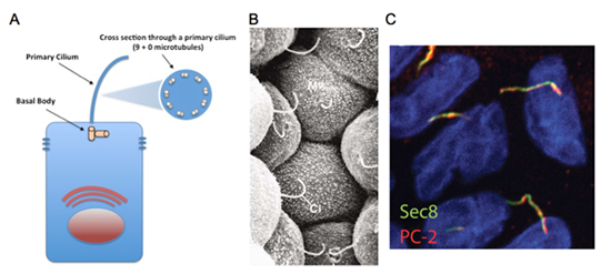
Cysts and tubules are primary building blocks of the kidney, and defects in cystogenesis or tubulogenesis during kidney development can result in a spectrum of pediatric and adult renal diseases. Relevant to our research, during kidney development, disruption in the formation of de novo epithelial cysts from pools of mesenchymal stem cells lead to various forms of congenital abnormalities such as kidney hypoplasia, dysplasia, or agenesis. On the other hand, following nephrogenesis, when the inhibitory signals that halt excessive cyst formation are disturbed, forms of renal disease such as polycystic kidney disease (PKD) can occur. Autosomal dominant PKD (ADPKD), which affects approximately 500,000 Americans and currently has no approved treatment, is the most common potentially lethal genetic disease. Every variation of human PKD has been attributed to mutations in genes important to the assembly or function of the primary cilium, a thin rod-like organelle on renal tubular epithelial cells which projects into the tubular lumen (See Figure 1).
Primary cilia have been found on the surface of most growth-arrested or differentiated mammalians cells, including a large variety of epithelial cells, endothelial cells, connective-tissue and muscle cells, as well as neurons and even embryonic stem cells. Although the presence of primary cilia has been noted for over a hundred years, its biological function was a mystery. Due to an exploding volume of research in just the last decade, it is now believed the primary cilium acts as a "sensory antenna" to relay signals to the cell based on the extracellular environment. These can include sensory clues such as mechanical stimulation (bending or rotation), chemosensation (detection of growth factors, morphogens, etc.), osmolarity, light, temperature, and even gravitational pull. It is thought that primary cilia along the renal tubules are responsible for detecting fluid composition and flow dynamics, and when defective, the cells misinterpret this as a signal to dedifferentiate and proliferate, resulting in the formation of large pathogenic renal cysts. However, defects in primary cilia can affect other tissues as well, and have resulted in a new classification of human disorders termed "ciliopathies".
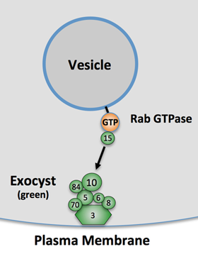
Currently, we have research projects focused both on the molecular mechanisms of de novo cyst formation, and on primary cilia assembly and signaling. We have found both of these cellular activities depend on the exocyst complex, a highly conserved eight-protein complex (see Figure 2), which is involved in targeted secretory vesicle transport. We are working hard to discover how cells regulate the exocyst subunits in order to direct polarized trafficking to specific locales such as primary cilia and the lumens of growing epithelial cysts. These discoveries may lay the groundwork for novel therapies for adult and pediatric renal diseases, and expand our knowledge of some of the basic cellular and molecular mechanisms by which the human body controls its development.
Affiliation: School of Medicine, Department of Anatomy, Biochemistry, and Physiology
Position: Assistant Professor
Phone: (808) 692-1009
Fax: (808) 692-5497
Email: jkenjih@hawaii.edu
Address: 651 Ilalo St., MEB 307R, Honolulu, HI 96813
Go to Jason Higa's webpage
Affiliation: School of Medicine, Department of Anatomy, Biochemistry, and Physiology
Position: Professor
Phone: (808) 692-1442
Fax: (808) 692-1951
Email: lozanoff@hawaii.edu
Address: 651 Ilalo St., BSB 119A, Honolulu, HI 96813
Go to Scott Lozanoff's webpage
 Yusuke Marikawa, PhD
Affiliation: School of Medicine, Institute for Biogenesis Research
Position: Professor
Degree: PhD (Kyoto University, Japan)
Phone: (808) 692-1411
Fax: (808) 692-1962
Email: marikawa@hawaii.edu
Address: 651 Ilalo St., BSB 163A, Honolulu, HI 96813
Axis Specification and Germ Layer Formation in Mammalian Embryos.
Differentiation and Morphogenesis in Pluripotent Stem Cells.
Stem Cell-Based Detection of Teratogens.
How can a single cell, like the fertilized egg, transform into a complex architecture, like our body? With respect to this biggest question of Developmental Biology, my lab is currently conducting research projects under the following two main themes:
"Axis Specification and Germ Layer Formation in Mammalian Embryos"
Soon after implantation, the embryo of human or mouse is a single layer of epithelial tissue, called the epiblast. Later, one edge of the epiblast forms the structure called the primitive streak, from which cells migrate away to form new tissue layers. This event, i.e., the formation of the primary germ layers (ectoderm, mesoderm, and endoderm), is the first step towards the generation of complex body patterns. Furthermore, the primitive streak formation is the first morphological landmark of body axis specification, as it establishes the caudal end of the future body.
Activation of Wnt/beta-catenin signaling, an evolutionary conserved signal transduction machinery, is the key to the formation of the primitive streak in the epiblast. The specific questions that are addressed in my current projects are:
1) How Wnt/beta-catenin signaling is activated in a localized region of the epiblast?
2) What genes are turned on by Wnt/beta-catenin signaling in the primitive streak?
3) How can cells in the primitive streak transform from epithelial into migratory state?
"Differentiation and Morphogenesis in Pluripotent Stem Cells"
Embryonic stem (ES) cells and induced pluripotent stem (iPS) cells can be maintained and propagated in a culture dish as undifferentiated cell populations, which are very similar to early embryonic cells before germ layer formation. Because studies of mammalian embryos are often hindered by the unique reproductive mode of mammals (i.e., embryos normally develop in the uterus), pluripotent stem cells, like ES and iPS cells, can serve as great in vitro models, which can be experimentally manipulated more easily. Furthermore, these pluripotent stem cells offer tremendous promise for the future regenerative medicine, as they can be used to derive various types of functional tissues in vitro, and those stem cell-derived tissues may be used for tissue replacement therapy and drug screening.
The goal of my lab is to elucidate molecular and cellular mechanisms of differentiation (e.g., of mesoderm and endoderm tissues) and morphogenesis (e.g., of tissue elongation along the body axis) using mouse and human pluripotent stem cells.
 Takashi Matsui
Affiliation: School of Medicine, Dept. of Anatomy, Biochemistry and Physiology, Center for Cardiovascular Research
Position: Professor and Chair
Degree: MD and PhD (Jikei Univ Sch of Med, JAPAN)
Phone: (808) 692-1554
Fax: (808) 692-1973
Email: tmatsui@hawaii.edu
Address: 651 Ilalo St., BSB 110, Honolulu, HI 96813
Go to Takashi Matsui's webpage
The role of mammalian target of rapamycin (mTOR) in the heart, Cardiac cell signaling controlling cell survival, Diabetic hearts, Ferroptosis
Our research is focused on the insulin signaling pathway in cardiomyocytes, especially cardioprotective effects against pathological settings such as ischemia. Our interests center around the role of the mechanistic target of rapamycin (mTOR), which is intimately related to the insulin/phosphatidylinositol 3-kinase (PI3K)/Akt signal transduction pathway. In order to investigate the role of mTOR in the heart, we utilize a variety of in vitro, in vivo, and ex vivo models of heart failure with genetically manipulated models of mTOR such as transgenic and knockout mice. We have reported that the role of cardiac mTOR in prevention of cardiomyocyte cell death that arises from myocardial infarction and cardiac hypertrophy, apparent risk factors for heart failure.
Diabetes is an independent risk factor for both heart failure and ischemic heart disease. Because of the important role of mTOR in insulin signaling, we have been working to determine the role of mTOR in diabetic hearts, and exploring the mTOR signaling pathway as a novel therapeutic target for treatment of heart failure in diabetes.
We recently demonstrated that mTOR is necessary and sufficient for cardiomyocyte protection against iron-mediated cell death that includes excessive iron-induced cell death and ferroptosis that is an iron-dependent form of regulated cell death. This is the first report that ferroptosis is a significant type of cell death in cardiomyocytes. We are currently defining the pathophysiological role of ferroptosis in cardiac diseases such as acute myocardial infarction and heart failure.
 Alika K. Maunakea, Ph.D.
Affiliation: YIBR/ABP
Position: Professor
Graduate Faculty: Molecular Biosciences and Bioengineering (MBBE)
Developmental and Reproductive Biology (DRB)
Cell and Molecular Biology (CMB)
Neuroscience Specialization Program
Phone: (808) 692-1048
Fax: (808) 692-1255
Email: amaunake@hawaii.edu
Address: 651 Ilalo St., Room 222K, Honolulu, HI 96813
Go to Alika Maunakea's webpage
 Stefan Moisyadi, PhD
Affiliation: School of Medicine, Institute for Biogenesis Research
Position: Associate Professor
Graduate Faculty: Developmental and Reproductive Biology (DRB)
Cell and Molecular Biology (CMB)
Phone: (808) 956-3118
Fax: (808) 956-7316
Email: moisyadi@hawaii.edu
Address: IBR, 1960 East-West Rd. Room E108, Honolulu, HI 96822
Affiliation: School of Medicine, Anatomy, Biochemistry & Physiology
Position: Assistant Professor
Phone: (808) 692-1422
Fax: (808) 692-1951
Email: polgar@hawaii.edu
Address: 651 Ilalo St., BSB 110, Honolulu, HI 96813
Go to Noemi Polgar's webpage
 Kathryn J. Schunke
Affiliation: Department of Anatomy, Biochemistry and Physiology, Center for Cardiovascular Research, Diabetes Research Center, John A. Burns School of Medicine
Position: Assistant Professor
Degree: PhD
Phone: (808) 692-1565
Email: kschunke@hawaii.edu
Address: 651 Ilalo St., BSB 211, Honolulu, HI 96813
 Johann Urschitz, PhD
Affiliation: Institute for Biogenesis Research Department of Anatomy,
Biochemistry and Physiology, John A. Burns School of Medicine
Position: Associate Professor
Graduate Faculty: Developmental and Reproductive Biology (DRB)
Cell and Molecular Biology (CMB)
Phone: (808) 956-7417
Fax: (808) 956-7316
Email: johann@hawaii.edu
Address: IBR, 1960 East-West Rd., Room E112, Honolulu, HI 96822
 Monika A. Ward, PhD
Affiliation: Institute for Biogenesis Research Department of Anatomy, Biochemistry and Physiology, John A. Burns School of Medicine
Graduate Faculty: Chair, Developmental and Reproductive Biology (DRB)
Cell and Molecular Biology (CMB)
Phone: (808) 956-0779
Fax: (808) 956-7316
Email: mward@hawaii.edu
Address: IBR, 1960 East-West Rd., Room E104, Honolulu, HI 96822
Go to Monika Ward's webpage
Sperm Genetics and Function in Fertilization
Assisted reproduction enables achieving fertilization when normal conception does not occur due to a variety of gamete defects. Intracytoplasmic sperm injection (ICSI) is the injection of a single spermatozoon directly into the cytoplasm of an oocyte using an injection pipette. This technique has been applied successfully in the treatment of infertile couples and is now widely used as a method of human assisted reproduction (ART). In addition to its obvious role in overcoming the infertility, ICSI is also an excellent tool allowing exploring new venues of reproductive biology. It provides a unique opportunity to examine the mechanisms underlying male infertility by looking into the reproductive potentials of individual spermatozoa.
The primary research interest of the lab is to study sperm genetics and function in fertilization in the context of assisted reproduction, utilizing advanced techniques of gamete and embryo micromanipulation combined with cytogenetic and molecular biology techniques, in a mouse model. The overall goal is to explore how the 'genetic composition' of sperm translates on its function in fertilization.
The ongoing projects involve:
(1) Reproducing subfertile and infertile mice with phenotypes that mimic various human male infertility syndromes to test for ART effects;
(2) Studying sperm DNA damage, its origin and consequences for fertilization and embryo development;
(3) Examining the function of the Y chromosome encoded genes in male fertility;
(4) Identifying and characterizing the mechanism of action of the Y chromosome encoded susceptibility locus mechanistically relevant to multiple sclerosis.

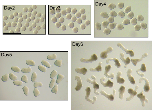
Figure 1. Teratozoospermia is a fertility defect expressed as an increased proportion of morphologically abnormal sperm that affects both humans and mice. Morphologically abnormal sperm (A) are often unable to fertilize oocytes rendering males infertile. Infertility can be overcome with intracytoplasmic sperm injection (ICSI), a method that allows injecting a spermatozoon directly into the oocyte using an injection pipette (B). The resulting embryos can be tested for their potential to develop in vitro from zygote to blastocyst stage (C), their genetic integrity evidenced by analysis of paternal or maternal chromosome complement in the zygote (D) and their potential to develop in vivo to live offspring after embryo transfer (E). Such analyses allow testing sperm function in infertile males with teratozoospermia, and can also be applied to other infertility defects.
 W. Steven Ward, PhD
Affiliation: Institute for Biogenesis Research Department of Anatomy, Biochemistry and Physiology, John A. Burns School of Medicine
Graduate Faculty: Developmental and Reproductive Biology (DRB)
Cell and Molecular Biology (CMB)
Phone: (808) 956-5189
Fax: (808) 956-7316
Email: wward@hawaii.edu
Address: IBR, 1960 East-West Rd., Room E108, Honolulu, HI 96822
Go to Steven Ward's webpage
The function and structure of mammalian sperm chromatin
Our research is focused on the structure of mammalian sperm chromatin and how this is related to function. The main hypothesis that we have tested is that the sperm cell provided the newly developing embryo with more than just the genetic code in the DNA sequence; it also provides a three dimensional organization of the DNA that provides crucial information as to how to use the father's genetic code.
DNA is packaged very densely in the sperm nucleus in a manner that is different from any other cell type. Most of the histones are replaced by protamines, and the DNA is crystallized into dense toroids with roughly 50,000 bp of DNA, each. Protamine condensation protects the sperm DNA from damage from external insults, and prevents transcription or DNA replication from occurring. We have shown that one structural feature present in all other somatic cells is also retained in sperm chromatin – the organization of DNA into loop domains attached at their bases to the nuclear matrix. This organization is crucial to two aspects of sperm chromatin function. Shortly after fertilization, the protamines are removed from the sperm DNA and the chromatin is repackaged with histones. The DNA is then replicated, and we have demonstrated that this DNA synthesis requires the loop domain organization to be intact. On the other hand, spermatozoa have the ability to digest their own DNA through an apoptotic-like process in which the sperm DNA is degraded. This degradation occurs on the nuclear matrix.
Our current research efforts are focused on understanding how sperm chromatin structure is related to the events that occur shortly after fertilization and how the DNA packaging in the sperm cell contributes to embryonic development.

Figure 1: Sperm DNA Loop Domain Organization is Required for the First Round of DNA Synthesis in the One Cell Embryo.
When normal mouse sperm heads are injected into oocytes, male (m) and female (f) pronuclei form, and DNA is replicated in each pronucleus (B). When the protamines are removed, and the only chromatin structure that is left is DNA loop domain organization, DNA replication still occurs (E). If some of the loop organization is removed by restriction endonuclease digestion, DNA replication still occurs (H). DNA alone, injected into the oocyte, does not result in DNA replication (K). Note that in this case, the female pronucleus still replicates its DNA.

Figure 2: (A) We have published evidence that each protamine toroid in the sperm chromatin is equivalent to a single DNA loop domain. (B) We have shown that when spermatozoa are induced to break their DNA at the nuclear matrix, they cannot support embryogenesis. When these sperm are injected into oocytes, they form normal pronuclei, but when DNA replication begins, the paternal DNA is degraded. One the other hand, when the loop DNA not directly associated with the nuclear matrix is digested with restriction endonucleses, spermatozoa can still support DNA replication. These data suggest that it is the association of DNA with the sperm nuclear matrix that is important for DNA replication of the paternal genome.
 Yukiko Yamazaki, PhD
Affiliation: Institute for Biogenesis Research Department of Anatomy, Biochemistry and Physiology, John A. Burns School of Medicine
Graduate Faculty: Developmental and Reproductive Biology (DRB)
Cell and Molecular Biology (CMB)
Phone: (808) 692-1416
Fax: (808) 692-1962
Email: yyamazak@hawaii.edu
Address: 651 Ilalo St., BSB 163-3 Honolulu, HI 96813
Go to Yukiko Yamazaki's webpage
Mechanisms of Sex Differentiation in Fetal Germ Cells.
Effects of Assisted Reproductive Technologies (ART) in Mammalian Development.
My primary research interest is the mechanisms of sex differentiation in fetal germ cells. In mammals, primordial germ cells (PGCs) are the precursors of sperm and oocytes. The sex-specific development of PGCs as sperm or oocytes is initiated by cues provided by the gonadal environment. After an initial sex differentiation event in the gonads, PGCs in a female gonad immediately enter meiosis, thereby committing oogenesis. On the other hand, PGCs in a male gonad do not enter meiosis until puberty. Instead, they first enter mitotic arrest and maintain G0 stage until after birth. Recent findings reveal that the sex-specific timing of meiosis entry is regulated by retinoic acid (RA) and RA degenerating enzyme in the gonads. Our goal is to elucidate how the molecular environment regulates the sex differentiation of fetal germ cells. Using a germ-cell culture system, we have examined mechanisms of entry into meiosis or mitotic arrest in the fetal germ cells. Besides meiosis, another unique event of the germ cells after sex differentiation is epigenetic reprogramming of genomic imprinting. Our group has examined how this epigenetic reprogramming of genomic imprinting is regulated to establish sex-specific imprints in the germ cells.
I am also interested in the effects of Assisted Reproductive Technologies (ART) in mammalian development. In human being, the majority of children conceived through ART, such as in vitro fertilization (IVF) and intracytoplasmic sperm injection (ICSI), appear healthy; however, metabolic abnormalities have been reported. As an animal model, we have studied the effects of these techniques on obesity and the epigenetic status of imprinted and non-imprinted genes in mouse offspring (collaborations).
 Yiqiang Zhang
Affiliation: JABSOM, Center for Cardiovascular Research
Phone: (808) 692-1480
Fax: (808) 692-1973
Email: yiqiang.zhang@hawaii.edu
Address: 651 Ilalo St., BSB-311D, Honolulu, HI 96813
Go to Yiqiang Zhang's webpage
Integrative Cellular Molecular Cardiobiology and Heart Regeneration
We study systems molecular regulations in cardiac growth and regeneration, stem cell and cardiomyocyte biology, and electrophysiology, using innovations in transgenic models, multi-omics (e.g., epigenetics, transcriptomics) and bioinformatics, functional physiology, and bioengineering.
INTRODUCTION: Our multi-disciplinary research covers cardiovascular molecular and cell biology and integrative pathophysiology and multi-omics sciences. We study cardiac and stem cell biology and heart regeneration, cardiac growth and disease mechanisms, cell cycle control, epigenetics and functional genomics, heart failure therapies, and electrophysiology and arrhythmias. By obtaining a better, in-depth understanding of mechanisms underlying endogenous heart growth and regeneration, as well as exogenous cardiac regeneration by innovative approaches in stem cell biology and bioengineering, we hope to develop novel and effective therapeutics to treat congestive and congenital heart diseases.

OVERVIEW: Heart cell hemostasis and regeneration undergo dynamic changes during development and in disease processes. Understanding the molecular and genetic pathways regulating cell lineage and functions is a crucial step in treating congenital and congestive heart diseases such as heart failure. Mammalian cardiomyocytes rapidly enter into a quiescent cell cycle state shortly after birth. Dr. Zhang's group first demonstrated that adult cardiomyocytes (ACMs) retain substantial cellular plasticity, capable of dedifferentiation, and can become more primitive to re-enter the cell cycle to proliferate, contributing to the endogenous myocardial regeneration in post-injury hearts, albeit at a low level. In addition, cardiomyocytes derived from pluripotent stem cells are great models and unlimited resources for exogenous heart regeneration. Still, molecular epigenetic regulations on cardiac cell biology and functions remain largely undetermined. An integrative system investigation in cell and molecular cardiobiology will reshape the future of preventing and treating heart disease.
GOALS AND APPROACHES: With the ultimate goal of treating degenerative heart diseases by promoting both endogenous and exogenous cardiac regeneration, the Zhang lab is working to determine the integrative cardiac and non-cardiac cellular processes and molecular pathways regulating cardiomyocyte differentiation, maturation, dedifferentiation, and cell cycle activities. His team uses state-of-the-art transgenic and reporter cells and animal models bridging to human health, cutting-edge multi-omics and bioinformatics approaches, together with advanced cellular, molecular, and bioengineering technologies.
CURRENT RESEARCH THEMES
Cardiomyocyte Growth, Dedifferentiation, and Cell Cycle Regulation
Based upon our early work in cardiomyocyte dedifferentiation, we have recently developed new multi-reporter transgenic mouse models for rigorous cardiomyocyte lineage tracing and real-time maturity (versus dedifferentiation) visualization (Figure 1). We continue studying molecular regulations of endogenous myocardial regeneration, namely cardiomyocyte dedifferentiation followed by proliferation, in injured (e.g., infarcted and hypertrophic) hearts. We use multi-disciplinary approaches, including high-throughput single-cell imaging, massive parallel single-nucleus RNA-seq and ATAC-seq, DNA methylome, and integrative cellular, molecular, and functional physiological analyses. We also use cell cycle-specific reporter systems and patient cardiac tissues to study heart cell hemostasis and mechanisms of endogenous myocyte renewal and regeneration.
Stem Cell and Cardiac Biology, Bioengineering, Cardiac Physiology, and Heart Regeneration
Pluripotent stem cells (e.g., induced pluripotent stem cells/iPSCs, or embryonic stem cells/ESCs) are the unique models used in cardiac development and heart regeneration research. We study cardiomyocyte differentiation, growth (maturation versus dedifferentiation), cell cycle (proliferation), cellular physiology, and how these processes are modulated by cellular cues such as bioengineered, nanopatterned surfaces (Figure 2), and their underlying molecular/epigenetic mechanisms. The overarching goals of our projects are to dissect molecular mechanisms regulating these multi-faceted processes in stem cells and heart cells and to generate important targets to enhance exogenous myocardial regeneration using cell therapies.
Integrative Functional Multi-Omics in Heart Diseases
We apply large-scale multi-omic approaches to discover and translate knowledge of treating cardiovascular diseases (Figure 3). By taking advantage of novel transgenic models for cell lineage, cell cycle, and specific molecular expression, and using both animal models and human biopsies, we study the integrative transcriptomic and epigenomic regulations (e.g., chromatin accessibility by ATAC-seq, histone and DNA modifications, and miRNA) in cardiac development and disease remodeling. Our systematic functional genomic work on global heart cell populations has the potential for comprehensive, in-depth knowledge and novel discoveries critical in treating heart failure and other cardiovascular diseases.

Figure 1: Modes of endogenous myocardial regeneration revealed in multi-reporter transgenic models for cardiomyocyte lineage tracking and maturity (vs dedifferentiation) reporting. Single-cell imaging using ImageStream revealed significantly increased BrdU+ cycling pre-existing myocytes in post-infarct hearts compared to sham, and that increase was preferentially in dedifferentiated BFPlow CMs.

Figure 2: Cardiac and Stem Cell Biology and Electrophysiology, and the roles of extracellular cues on myocyte maturity and functions revealed by assays using microelectrode arrays.

Figure 3: System cardiac genomics. A, t-SNE plot showing cell populations in normal and post-infarct triple-transgenic mouse hearts revealed by massive parallel single-nucleus RNA-seq (snRNA-seq) analysis. B, Integrative transcriptomic and DNA methylomic analysis of differentially expressed (DEGs) or methylated genes (DMGs) in dedifferentiated cardiomyocytes.
 Peter R. Hoffmann, PhD, MSPH
peterrh@hawaii.edu | (808) 692-1510
Full Member, Cancer Biology Program, University of Hawaiʻi Cancer Center
Academic Appointment(s): Professor, Cell and Molecular Biology Department, John A. Burns School of Medicine, University of Hawaiʻi at Mānoa
Degree(s): PhD, Immunology and Microbiology, University of Colorado
MSPH, Public Health, University of Hawaiʻi at Mānoa
 Thomas Huang, PhD
Associate Professor
Affiliation: JABSOM, Obstetrics, Gynecology and Women's Health
Email: huangt@hawaii.edu
Phone: (808) 203-6533
Fax: (808) 955-2174
Address: Kapiolani Med Ctr
Go to Thomas Huang's webpage
 Olivier Le Saux, Ph.D.
Professor and Chair, Department of Cell and Molecular Biology (CMB)
Area of Expertise: Skin and cardiovascular diseases, ectopic calcification
Email: lesaux@hawaii.edu
Phone: (808) 692-1504
Go to CMB Faculty webpage
Assistant Professor
Affiliation: JABSOM, Obstetrics, Gynecology and Women's Health
Go to Corrie Miller's webpage
 Birendra Mishra PhD, MS, DVM
Assistant Professor
Department of Human Nutrition, Food & Animal Sciences, College of Tropical Agriculture and Human Resources – University of Hawai‘i at Mānoa
Email: bmishra@hawaii.edu
Phone: (808) 956-7021
Fax: (808) 956-4024
Principal Investigator
Phone: (808) 691-7902
Fax: (808) 691-7939
miortega@queens.org
Go to CMB Faculty webpage
 Jesse B. Owens, PhD
Affiliation: Institute for Biogenesis Research
Department of Anatomy, Biochemistry and Physiology
John A. Burns School of Medicine
Graduate Faculty: Developmental and Reproductive Biology (DRB)
Cell and Molecular Biology (CMB)
Phone: (808) 956-4828
Fax: (808) 956-7316
Email: jbowens@hawaii.edu
Address: IBR, 1960 East-West Rd, Room E108, Honolulu, HI 96822
Go to CMB Faculty webpage
 Michelle Tallquist, PhD
Affiliation: JABSOM, Center for Cardiovascular Research
Position: Professor
Degree: PhD (Immunology, Mayo Clinic and Foundation)
Phone: (808) 692 1579
Fax: (808) 692 1973
Email: michelle.tallquist@hawaii.edu
Address: BSB 311E, 651 Ilalo Street, Honolulu HI, 96813
 Jinzeng Yang, PhD
Affiliation: Department of Human Nutrition, Food and Animal Sciences
Position: Associate Professor
Degree: PhD (University of Alberta, Canada)
Phone: (808) 956 6073
Fax: (808) 956 4024
Email: jinzeng@hawaii.edu
Address: 1955 East West Road, Rm 216, Honolulu, HI 96822
Go to Jinzeng yang's webpage
Affiliation: JABSOM, Obstetrics, Gynecology and Women's Health
Professor
Go to Claire Wright's webpage
Affiliation: Geriatric Medicine
Position: Professor
Phone: (808) 523-8461
Fax: (808) 528-1897
Email: willcox@hawaii.edu
Address: Kuakini Med Ctr HPM-9
Go to Bradley Wilcox's webpage